ANATOMY OF THE EYE
The eye is that wonderful organ of sense that allows us to see and its structure closely resembles the one of a camera.
Actually, we have created the camera "copying" what nature had already invented.
When we fix an object, the light that comes from it enters our eyes, passes through a series of lenses, that are in sequence: the cornea, the aqueous humour, the crystalline and the vitreous which correspond in the camera to the lenses of the objective, and then it goes "to impress" the retina (film).
The retina, excited by the light that hits it, transmits the information to the "director", the brain, through a electrical wire, the optical nerve.
The brain studies and exploits the visual information, taking advantage of it in order to decide the behaviour and the reactions of the whole organism.
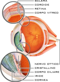
Sclera
The sclera, the so-called “white of the eye”, is the outermost and sturdiest membrane of the eye and it is composed of fibrous tissue. In the front part of the eye, where it becomes transparent and it is curved as like the glass of a watch, it takes the name of cornea.
Cornea It's the first natural lens the incoming light encounters. It is essential that it is transparent and of spherical shape. A not perfectly spherical cornea can cause astigmatism.
The cornea is a transparent membrane without blood vessels but extremely rich in nervous fibres. It is kept continuously wet by a lacrimal film adhering to its surface. The interface between the corneal surface and lacrimal film constitutes the most powerful convergent lens on the human eye. The cornea has a thickness of approximately 1 millimetre and is composed (from the outer surface) of 5 layers: The adult cornea is only about 1/2 millimetre thick and is comprised of 5 layers: 1) epithelium, 2) Bowman's membrane, 3) stroma, 4) Descemet's membrane and 5) the endothelium. The stability of the lacrimal film and the transparency of the cornea are essential for vision. Behind the cornea is the anterior chamber, filled with a thin, watery fluid called aqueous humour. Traumas or infections can cause the formation of permanent opacities of this natural lens, which, losing its transparency, will limit the vision. Iris
The iris is the part front of the uvea that gives the colour to our eyes and encircles a smallcentral hole of variable amplitude between 2 to 8 millimetres. The pupil is composed of a stroma (interdigitated sheets of collagen material), vessels and two muscles: the dilator muscle (that widens the pupil size) and the sphincter muscle (that narrows the pupil size). Its colour can be fair (from blue to green) or dark (from brown to black) but in truth its coloration depends both on the amount of pigmentation and on optical phenomena of reflection and diffraction of the light in the stroma.
In an iris with little pigmentation, light passes through the deep layers where it is reflected assuming a light colour. On the other end, in a pigment-rich iris, light is not able to reach the deep layers and it is not reflected nor refracted.
The iris encircles the pupil, which increases in size or shrinks according to amount of light reaching it, and therefore acting just like the diaphragm of a camera that regulates how much light can reach the film. Behind the iris is the crystalline lens.
Cristalline
The crystalline is a convergent lens of biconvex shape that focuses the rays of light on the retina. While in a camera the photographer focuses the image varying the focal distance between lens and film, in the eye the distance between the crystalline lens and the retina remain fixed.
The eye focuses objects at various distances with a different strategy: the crystalline lens has the ability to continuously modify its shape and to vary its curvature so as to increase or to diminish its power of convergence. This dynamic process, known as accommodation, is controlled by a ring of muscular fibres located around to the crystalline lens and called the ciliary body. Thus, when the eye is focusing a distant object, the crystalline lens flattens and diminishes its curvature. When the eye is focusing a near object, the crystalline lens becomes more convex and increases its curvature. Aging induces in both the crystalline lens and the ciliary body a loss of the power of accommodation, making us long-sighted and unable to read from a distance of 30 cm. In this case, it is necessary to turn to the artificial optical correction offered by bifocal or multifocal lenses. Moreover, the appearance of opacities in the crystalline lens (cataract), especially if in the centre of the field of view, can disturb the vision. To restore optical transparency, the cataract can be surgically operated and replaced with a small artificial lens Vitreous
The vitreous is a clear jelly-like substance contained in the vitreal cavity that, filling up the space comprised between the crystalline lens and the retina, maintains the spherical shape of the eyeball. Its transparency is important for a clear vision at all distances. Opacity of the vitreous as a consequence of inflammatory or hemorrhagic processes can seriously compromise the visual ability. With aging, the vitreous loses its consistency, gets detached and fluctuates in the vitreal cavity. One symptom of a severe separation of the vitreous is the appearance of mobile bodies often associated with flashes. In these cases, an examination of the bottom of the eye in order to search for possible retinal breaches that could in some cases lead to retina detachment is imperative.
Retina
The retina, analogous of the photographic film, covers the internal surface of the ocular globe. It appears as a thin transparent membrane, subdivided in two areas: a central area - the macula - containing the fovea and rich of cones and a peripheral area, where the rods prevail, used for peripheral and nocturnal vision.
After having crossed the cornea, the anterior chamber, the pupil, the crystalline lens and the vitreous, the rays of light converge on the retina and in particular in that very small area called fovea: a highly specialized structure that supervises, in conditions of high brightness, to the maximum visual acuity of close and distant objects, the perception of colours and the sensitivity to contrast.
The more complex mechanisms of vision take place in the retina. The light passes through the entire thickness of the retina (see figure) and immediately hits the photoreceptors, cones and rods, which constitute the outermost part of the retina in contact with the layer of cells of the retinal pigment epithelium (RPE). The integrity of the RPE is essential in order to mediate the metabolic exchanges between photoreceptors and the underlying choroid.
The photochemical processes of the vision can be outlined in two phases:
Photochemical reaction: the light is absorbed by photosensitive pigments (iodopsin in the cones, rodopsin in the rods) that decompose and give origin to a chemical reaction that converts a luminous signal into an electrical nervous impulse.
Transmission of the impulse: the electrical nervous impulse is transmitted to the bipolar and ganglion cells and, through the optical nerve, leaves the eye and arrives to the visual centre of the brain.
Choroid
Below the retina is the choroid. The choroid is the posterior part of the uvea. It is the vascular membrane of the eye. It consists of layers (a layer of large choroidal vessels and a layer of coriocapillary vessels) and of the Bruch membrane in contact with the RPE. Its function is to nourish and to oxygenate the RPE, the outermost retinal layers (in particular the photoreceptors) through the Bruch membrane, as well as contributing to the blood supply of the optical nerve.
Optical nerve
The optical nerve consists of more than a million nervous fibres that originate from the ganglion cells of the retina. Its function is to connect the eye to the visual centre of the brain, called occipital visual cortex. Here the incoming nervous impulses are processed and transformed into the visual perception of an image. The connection retina-brain through the optical nerve is so tight that the vision is considered a very complex phenomenon that happens mainly at cerebral level.